Loading
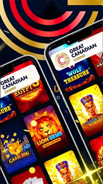
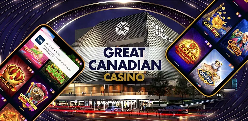
Great Canadian Casino
Great Canadian Casino inc.![]()
Contains ads · In-app purchases
of 9 MB
5![]()
11624 Comments
9 MB
![]()
18+
500000
Installed
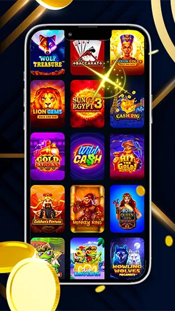
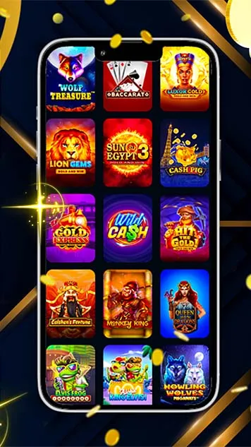
🍁Great Canadian Casino brings the vintage charm right to your phone! Enjoy exciting slot machines, vibrant visuals, and a welcome bonus of 100 FREE SPINS. Feel the thrill of Flamingo’s iconic style — no passport needed, just pure fun! 🍁
![]() This application does not share data with third parties
This application does not share data with third parties
![]() Data is encrypted during transmission
Data is encrypted during transmission
![]() This app not collect data
This app not collect data
![]() Data isn't encrypted
Data isn't encrypted

Great Canadian Casino
Rating and review
W
WinterFrost
Overall a solid casino game with engaging features. I recommend it!
Developer
Thank you for recommending us! We hope you continue to enjoy the game.172 people found this review helpful.
Was this information useful to you?
C
Cherry
The daily rewards are a nice touch! It keeps me coming back each day. Very engaging!
Developer
Thanks for noticing! We believe in rewarding our players for their loyalty.139 people found this review helpful.
Was this information useful to you?

A solid casino experience overall! From the variety of games to the engaging gameplay, it's a winner.
Developer
Thank you for your feedback! We're glad you had a solid experience.90 people found this review helpful.
Was this information useful to you?

Great graphics and engaging gameplay! I've had a lot of fun spinning the reels. Highly recommend this casino game!
Developer
Thank you for your kind words! We're delighted to hear you're enjoying the experience.112 people found this review helpful.
Was this information useful to you?

I love the variety of games available! There's always something new to try. Definitely a 5-star experience.
Developer
We appreciate your support! We're always working on adding more exciting games.154 people found this review helpful.
Was this information useful to you?
P
Petal
Amazing sound effects! They really enhance the atmosphere of the game. I'll keep playing for sure!
Developer
We're so happy you love the sound effects! Enjoy your gaming experience!159 people found this review helpful.
Was this information useful to you?
A
AuroraBorealis
I had a great time playing the slots! The graphics are stunning and the payouts are generous. Will definitely be back for more!
Developer
Thank you for your wonderful feedback! We're thrilled you enjoyed the slots.111 people found this review helpful.
Was this information useful to you?

The game runs smoothly with no glitches. A reliable option for casino fun!
Developer
We're happy to hear the game runs smoothly for you! Thank you for playing.161 people found this review helpful.
Was this information useful to you?

Just played blackjack and it was thrilling! I appreciate the realistic gameplay and interactions.
Developer
Thank you for your review! We're glad you found the gameplay engaging.104 people found this review helpful.
Was this information useful to you?

Fun gameplay and stunning animations! I could play for hours without getting bored.
Developer
Happy to hear you're enjoying the visuals! We have more exciting features coming soon.94 people found this review helpful.
Was this information useful to you?

I love the variety of games available! The graphics are stunning and really immersive. Definitely a top-notch casino experience.
Developer
Thank you for your positive feedback! We're glad you enjoy our graphics and game selection.83 people found this review helpful.
Was this information useful to you?
F
FlamePrincess
Fun game with lots of chances to win! I appreciate the daily rewards.
Developer
We're glad to hear you're enjoying the rewards system! Keep playing!177 people found this review helpful.
Was this information useful to you?
S
Sunburst
Incredible jackpots! I've won more than I expected. This game is definitely worth playing!
Developer
We're excited to hear about your wins! Best of luck with your future games.95 people found this review helpful.
Was this information useful to you?

Great casino game, but I wish they had more slot options. Overall, a nice experience.
Developer
Thank you for your input! We're constantly looking to expand our offerings.137 people found this review helpful.
Was this information useful to you?

I appreciate the fair play and transparency. It's refreshing to see a game that values its players.
Developer
Thank you for your trust! We strive for fairness and integrity in our gaming.135 people found this review helpful.
Was this information useful to you?
F
FairyDust
A fantastic casino game! The interface is user-friendly and very intuitive. I find myself playing for hours!
Developer
We're delighted to hear you find our interface easy to use! Enjoy your gaming experience!145 people found this review helpful.
Was this information useful to you?
H
Harmony
Excellent graphics and easy to understand rules. Perfect for beginners!
Developer
Thanks for the feedback! We strive to make our games accessible for everyone.129 people found this review helpful.
Was this information useful to you?
E
EmeraldHeart
Fantastic casino game! The jackpots are exciting, and I love the thrill of winning!
Developer
We appreciate your enthusiasm for the jackpots! Good luck with your next win!153 people found this review helpful.
Was this information useful to you?

This casino app is amazing! I love the bonuses and rewards system. Keep it up!
Developer
We're thrilled you love the bonuses! Thanks for playing!90 people found this review helpful.
Was this information useful to you?

Amazing experience overall! I found myself immersed in the gameplay.
Developer
We're thrilled to hear you had an amazing experience! Thanks for your support.164 people found this review helpful.
Was this information useful to you?
B
Blossom
Absolutely love this game! It really feels like I'm at a real casino. The ambiance is perfect!
Developer
Wow, thank you! We're working hard to replicate that authentic casino feel.158 people found this review helpful.
Was this information useful to you?

Love the community aspect! It's great to interact with other players while enjoying a game. Really fun!
Developer
We're so glad you enjoy the community feature! Happy gaming!95 people found this review helpful.
Was this information useful to you?
A
AuroraSky
Great game mechanics and smooth gameplay! I can't believe how realistic it feels. Highly recommended for anyone who loves casino games.
Developer
Thank you for recommending our game! We strive to create a realistic experience for our players.162 people found this review helpful.
Was this information useful to you?

This casino game is just what I needed. Smooth interface and great animations!
Developer
We're happy to hear you love the interface and animations! Enjoy playing!127 people found this review helpful.
Was this information useful to you?

The daily challenges keep things interesting! I really appreciate the effort put into this game. Great job!
Developer
Thank you for noticing! We're committed to keeping the game fresh.182 people found this review helpful.
Was this information useful to you?
R
RainbowDreamer
Great game! The graphics are stunning and really immersive. I've been enjoying it for hours.
Developer
Thank you for the amazing feedback! We're thrilled you're enjoying the graphics.160 people found this review helpful.
Was this information useful to you?
S
Starlight7
I love the variety of slot machines! Each game offers something unique and fun. Definitely a fun experience!
Developer
Thanks for your feedback! We're glad you enjoyed our variety of slots.116 people found this review helpful.
Was this information useful to you?

The live dealer games are a blast! The interaction makes it feel like I'm at a real casino. Highly addictive!
Developer
We're thrilled you enjoy our live dealer games! Thank you for your support.93 people found this review helpful.
Was this information useful to you?

Fantastic bonuses and rewards! I keep coming back for more. The gameplay is smooth and enjoyable.
Developer
We're thrilled you love the bonuses! Stay tuned for even more surprises.94 people found this review helpful.
Was this information useful to you?
L
LaceDove
Great experience overall! The slots are fun and rewarding. I can't stop playing them!
Developer
We're thrilled to hear that you're having a great time with our slots! Keep spinning!183 people found this review helpful.
Was this information useful to you?
Thank you for your feedback!


W
WinterFrostOverall a solid casino game with engaging features. I recommend it!
Developer
Thank you for recommending us! We hope you continue to enjoy the game.132 people found this review helpful.
Was this information useful to you?
C
CherryThe daily rewards are a nice touch! It keeps me coming back each day. Very engaging!
Developer
Thanks for noticing! We believe in rewarding our players for their loyalty.127 people found this review helpful.
Was this information useful to you?

EtherealA solid casino experience overall! From the variety of games to the engaging gameplay, it's a winner.
Developer
Thank you for your feedback! We're glad you had a solid experience.120 people found this review helpful.
Was this information useful to you?

EmeraldGreat graphics and engaging gameplay! I've had a lot of fun spinning the reels. Highly recommend this casino game!
Developer
Thank you for your kind words! We're delighted to hear you're enjoying the experience.102 people found this review helpful.
Was this information useful to you?

SunsetWhisperI love the variety of games available! There's always something new to try. Definitely a 5-star experience.
Developer
We appreciate your support! We're always working on adding more exciting games.115 people found this review helpful.
Was this information useful to you?
Show all review